



Your MRI or X-ray came back normal, but weeks on, your lower back or buttock pain is still there. It’s natural to wonder what the scan missed. Persistent pain after a normal scan is real, and it doesn’t mean the scan was wrong. Sometimes the source is simply a structure the scan wasn’t looking at, such as the sacroiliac joint.
Why doesn’t my MRI show my sacroiliac joint pain?
Sacroiliac (SI) joint pain often doesn’t show up on a standard lumbar MRI or X-ray because those scans assess the discs and lumbar spine, not the SI joint. Clinicians identify this functional problem by checking how the joint responds to a targeted local anaesthetic injection rather than how it looks on a picture. Researchers estimate that SI joint pain accounts for 15 to 30% of chronic low back pain, yet imaging alone pinpoints it in only a minority of cases.
TL;DR:
- SI joint pain, sometimes called SI joint dysfunction or SIJ pain, is thought to cause 15 to 30% of chronic low back pain.
- Common causes include pregnancy, asymmetric loading after an injury, spinal fusion surgery, and inflammatory conditions such as ankylosing spondylitis.
- Diagnosis combines a clinical history, provocative tests such as the FABER and thigh thrust tests, and a diagnostic block, since no single test settles it alone.
- SI joint pain can mimic sciatica or a slipped disc, but the underlying mechanism and the treatment pathway are different.
- Treatment usually moves from physiotherapy through image-guided injections to radiofrequency denervation for longer-lasting relief, once the diagnosis is clear.
What Is the Sacroiliac Joint, and What Does SI Joint Pain Feel Like?
The sacroiliac joints sit either side of the base of the spine, where the sacrum meets the ilium. Each one carries load between the upper body and the legs. It works every time you stand, walk or turn. Thick ligaments hold it stable, and only a little movement happens there, far less than in the lumbar spine above.
SI joint pain usually sits on one side, low down near the pelvis. It can spread into the buttock, groin or upper thigh. It often worsens when you stand up from sitting, climb stairs or roll over in bed.
A deep ache is more typical than a sharp pain. Many people search for “lower back and buttock pain one side” trying to describe exactly this pattern. Some get referred pain down the leg, which is often mistaken for sciatica. Read more about back pain and when to see a specialist.
When to Get Help Immediately
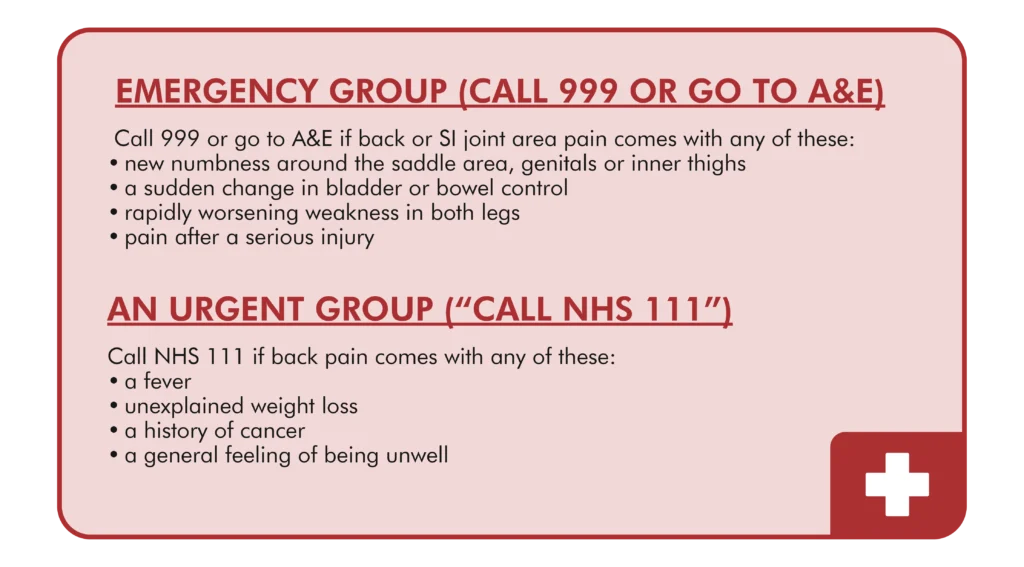
Most SI joint pain is not an emergency. It usually responds well to assessment and treatment over weeks rather than days. The exceptions are the red flags in the box above.
The emergency signs are new numbness, a bladder or bowel change, sudden weakness in both legs, or significant trauma. Any one of these means calling 999 or going to A&E, not waiting for a routine referral. A fever with back pain, unexplained weight loss or a cancer history still needs urgent same-day assessment, which NHS 111 or your doctor can arrange.
Why a ‘Normal’ Scan Doesn’t Rule Out Sacroiliac Joint Pain
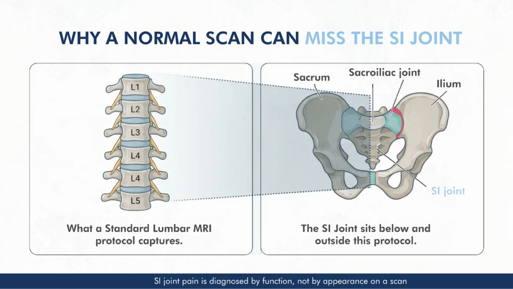
What a Standard Lumbar MRI Actually Looks At
A standard lumbar MRI checks the discs, the nerve roots and the lumbar spine, and it does that job well. Clinicians use it to examine the lumbar spine, not the sacroiliac joints. Parts of the joints may appear at the edge of the scan. However, clinicians sometimes need a dedicated SI joint protocol, for example when they suspect inflammatory sacroiliitis.
The 2025 international consensus guidelines describe sacroiliac joint complex pain as an underappreciated source of chronic low back pain. On imaging, the evidence for confirming it is unclear or negative.
Why the SI Joint Is Different
Unlike a disc problem, sacroiliac joint pain is defined by function rather than appearance. The joint can look unremarkable on a scan and still be the source of significant pain. That is because the picture rests on how it behaves under load and how it responds to a targeted injection. How it looks in cross-section matters far less.
When Imaging Does Help
Imaging still has a role. NICE’s guidance on imaging for low back pain sets out when a scan genuinely changes management. An X-ray or MRI can help rule out other causes, such as a fracture, an inflammatory condition or nerve compression.
So a normal scan does not close the door on SI joint pain. It simply means the scan did not target the SI joint.
What Causes Sacroiliac Joint Pain?
Pregnancy & Childbirth
Hormonal changes loosen the pelvic ligaments during pregnancy. As the added load passes through the joints, this can trigger pain that sometimes continues after birth. Clinicians often call this pelvic girdle pain.
Trauma & Asymmetric Loading
A fall, a road traffic collision, or years of uneven loading can all place extra strain on one sacroiliac joint. Uneven loading often comes from a leg-length difference, or from a physically one-sided occupation.
After Spinal Fusion Surgery
Lumbar spinal fusion changes how load passes through the pelvis, so the sacroiliac joint below the fused segment can take on extra strain over time. Pain can develop months or years later. The 2025 international consensus guidelines name this added loading and shear force as a potential contributor to SI joint pain.
Inflammatory Sacroiliitis
In younger patients especially, sacroiliac joint pain can be inflammatory rather than mechanical. Ankylosing spondylitis and related conditions cause sacroiliitis. This group needs a different diagnostic and treatment pathway from mechanical SI joint pain.
How Is Sacroiliac Joint Pain Actually Diagnosed?
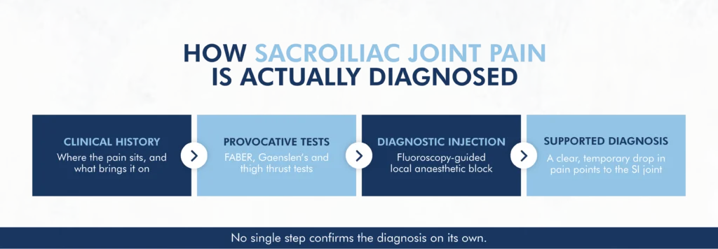
Clinical Examination & Provocative Tests
No single test identifies sacroiliac joint pain on its own. A pain specialist typically uses a cluster of provocative tests, including the FABER test, Gaenslen’s test and the thigh thrust test.
These sit alongside a detailed history of where the pain sits and what brings it on. A positive result across several of the tests raises suspicion, though it does not settle the diagnosis by itself.
The Diagnostic Injection: A Useful but Imperfect Test
A fluoroscopy-guided injection is often the most helpful single step. Under X-ray guidance, a small amount of local anaesthetic is placed into the joint. If your usual pain then drops clearly, even if only for a while, that supports the SI joint as the source.
It does not prove it on its own, though. False-positive and false-negative results can happen, so the result is read alongside your history and examination. A 2024 narrative review in Pain Practice describes this block as the current reference test, while making the same point: on its own, it is not infallible.
Sacroiliac Joint Pain vs Sciatica vs a Slipped Disc: How to Tell the Difference
Sciatica and a slipped disc typically involve compression or irritation of a nerve root. That produces pain running clearly down the leg, often below the knee. It sometimes comes with numbness, tingling or weakness in a set pattern.
Sacroiliac joint pain, by contrast, usually stays higher and more localised around the pelvis and buttock. It can still refer into the thigh, but rarely causes true numbness or weakness in the foot.
If you are unsure which pattern fits your pain, that uncertainty is itself a reason to get assessed, since treating the wrong structure only delays relief. Read our guide to sciatica and groin pain for the nerve-related picture.
Treatment Options That May Help
Treatment for sacroiliac joint pain usually follows a step-by-step path, much like chronic pain generally. You start with the least invasive option and move on only where it is needed.
Physiotherapy & Activity Modification
When clinicians suspect mechanical SI joint pain, they usually recommend targeted physiotherapy first. It focuses on pelvic stability and correcting asymmetric loading. Many people improve with this alone, particularly when pregnancy or a specific injury triggers the pain.
Image-Guided SI Joint Injections
When physiotherapy alone is not enough, clinicians may use an image-guided injection in two different ways. They agree on the purpose before the procedure.
A local-anaesthetic injection is part of the diagnostic picture, as above. A steroid injection is a treatment step that can give some patients short-term relief. Combining them in one injection can blur the diagnostic read, which is why the aim is agreed first.
Radiofrequency Denervation for Longer-Lasting Relief
Some patients respond to the diagnostic and prognostic blocks but still need longer-lasting relief. For them, radiofrequency denervation may be an option. It targets the small sensory nerves that supply the joint, in the posterior sacral network, commonly the L5 dorsal ramus and the S1 to S3 lateral branches.
This differs from radiofrequency treatment for a lumbar facet joint, which targets the medial branch nerves instead. That is why confirming the right structure first matters so much.
The SI joint has its own evidence base, separate from the lumbar spine or the neck, and the honest figure is more modest than for either. A randomised trial by Cohen and colleagues, published in Anesthesiology in 2008, followed 28 appropriately selected patients. It found that 57% still had at least 50% pain relief at 6 months.
Cooled radiofrequency techniques have reported relief in a similar 6-to-12-month range for those who respond. Results vary, though, and none of this is guaranteed.
How Dr Mehrez Diagnoses and Treats Sacroiliac Joint Pain
Dr Mehrez established a joint spinal and pain clinic and works in it alongside spinal surgeons. So a pelvic or lower-back pain presentation is looked at from both a pain-medicine and a spinal angle. It is not considered in isolation. He is registered with the General Medical Council, GMC 5171889, and holds the FRCA and FFPMRCA.
Where a diagnostic block or radiofrequency denervation is appropriate, these use image-guided, fluoroscopic technique. It is the same standard described on the radiofrequency treatment. For what a first appointment involves, our guide to what happens at a pain clinic walks through it. Independently published patient feedback, collected under the CMA’s private healthcare order, is on his PHIN profile.
Frequently Asked Questions
What does sacroiliac joint pain feel like?
A deep, one-sided ache low down near the pelvis, spreading into the buttock, groin or upper thigh. It often worsens with standing up from sitting, climbing stairs or rolling over in bed.
Why doesn’t sacroiliac joint pain show up on an MRI?
A standard lumbar MRI checks the discs and lumbar spine, not the sacroiliac joint. SI joint pain is a functional diagnosis. Clinicians identify it by checking how the joint responds to a targeted injection, not by looking at its appearance on a scan.
How is sacroiliac joint pain diagnosed?
Diagnosis combines a clinical history, provocative tests such as the FABER and thigh thrust tests, and a fluoroscopy-guided injection. A clear, temporary drop in pain after the injection supports the joint as the source. Your clinician considers this result alongside the rest of the picture, not on its own.
Can sacroiliac joint pain be cured?
Many people get substantial, lasting relief through physiotherapy, targeted injections and, where appropriate, radiofrequency denervation. Results vary, so ongoing management is a more honest description than a guaranteed cure.
What’s the difference between SI joint pain and sciatica?
Sciatica involves a compressed or irritated nerve root. The pain runs clearly down the leg, often with numbness or weakness in a set pattern. Sacroiliac joint pain usually stays higher, around the pelvis and buttock, and rarely causes true numbness in the foot.
Is sacroiliac joint pain common after pregnancy?
Hormonal changes loosen the pelvic ligaments during pregnancy. The added load through the joints can trigger pain that sometimes continues after birth, often called pelvic girdle pain.
How long does an SI joint injection take to work?
A diagnostic block often produces a noticeable, if temporary, drop in pain within minutes. That drop is part of what it tells us. A therapeutic steroid injection typically takes longer, often building over several days.
Do I need a GP referral to be assessed for SI joint pain?
Using insurance, you’ll usually need a GP referral to claim. Paying for yourself, it’s often not required, though this varies by consultant, so it’s worth checking when you book.
Can a chiropractor fix sacroiliac joint pain?
Some people get relief from manual therapy, particularly for mild symptoms. It is not a substitute for diagnosis, though. SI joint pain can also come from inflammatory conditions or nerve compression, which a clinical assessment would catch.
Does sacroiliac joint pain go away on its own?
Sometimes, especially where pregnancy or a minor injury set it off. Where the cause is ongoing asymmetric loading or an inflammatory condition, pain often persists without targeted treatment. There is no reliable way to tell which pattern applies without an assessment.
If persistent pain shows up higher in the spine instead, our guide to constant neck pain may help. It covers a similar pattern of red flags and diagnostic steps for the neck.
Where to See Dr Mehrez for SI Joint Assessment
Dr Mehrez sees patients for sacroiliac joint assessment at 107 Harley Street, London W1G 6AL. He also holds clinics at:
- The Saxon Clinic, Milton Keynes
- The Wellington Hospital, London NW8
- Three Shires Hospital, Northampton
- The Royal Buckinghamshire Hospital, Aylesbury
- Spire Harpenden Hospital
- The New Foscote Hospital, Banbury
Had a normal scan but the pain hasn’t gone away? Request a written second opinion, or ask about a short e-consultation first if you’d rather not book a full appointment yet.